Create Your First Project
Start adding your projects to your portfolio. Click on "Manage Projects" to get started
Heterochromatin Analysis in TEM Images
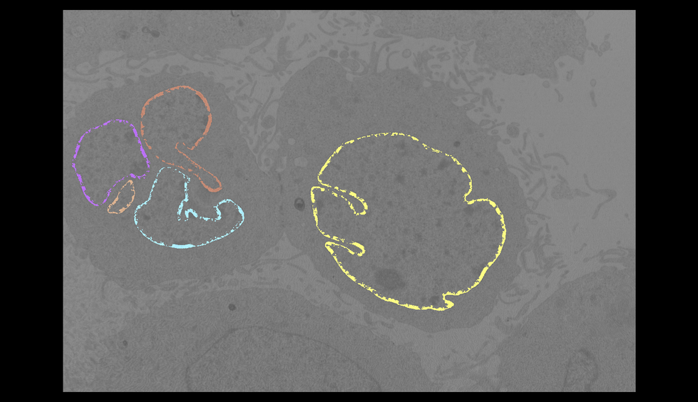
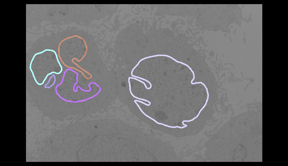
The goal of this project was to measure the amount of heterochromatin along the nuclear envelope in transmission electron microscope (TEM) images and quantify shape descriptors of the nuclei. Two of the major challenges of working with TEM data are that the counterstaining is not identical from section to section, and that the location of the section cannot be guaranteed to be the same from sample to sample (unless you are collecting serial TEM images for 3D reconstruction!). This means that direct measurements cannot be used (e.g., density of staining or raw area or perimeter measurements); instead, methods that provide normalized measurements are necessary.
To obtain normalized measurements of the heterochromatin, I generated a 25-pixel-wide band along the nuclear envelope, masked the heterochromatin in the nucleus, and then calculated the percentage of the band covered by heterochromatin.
For normalized measurements of the nuclear compartment, I calculated the circularity, eccentricity, solidity, and number of fragments to provide several options for comparing potential changes in the nucleus.
All steps of the analysis were carried out in Python using scikit-image, NumPy, and Pandas.


